What Is a Phyllodes Tumour?
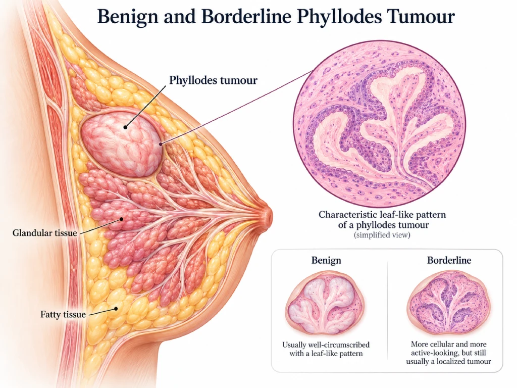
Phyllodes tumours are fibroepithelial tumours that arise from the connective tissue (stroma) of the breast. Most phyllodes tumours are benign, although some may be classified as borderline or malignant based on their microscopic features.
Benign and borderline phyllodes tumours are not breast cancers, but they can grow rapidly and may recur locally even after removal.
Phyllodes tumours can closely resemble fibroadenomas on clinical examination, ultrasound and mammography, making them difficult to distinguish before surgery.
Symptoms
- A painless breast lump
- A lump that increases in size over time
- Occasionally stretching of the skin if the tumour becomes large
Phyllodes tumours are usually mobile and well-defined on examination.
How Is It Diagnosed?
Assessment may include:
- Clinical breast examination
- Ultrasound and/or mammogram
- Core needle biopsy
In some cases, it may be difficult to distinguish a phyllodes tumour from a fibroadenoma with certainty, even on core biopsy.
The final diagnosis and classification (benign, borderline, or malignant) are usually confirmed after the lump has been surgically removed and examined under a microscope.
What Is the Difference Between Benign and Borderline Phyllodes Tumours?
Benign phyllodes tumours have lower risk features under the microscope and generally behave in a less aggressive manner.
Borderline phyllodes tumours have some features that suggest a higher risk of recurrence compared to benign tumours, but they are not considered frankly malignant.
Both benign and borderline phyllodes tumours are usually treated with surgery.
Treatment
Surgical removal is generally recommended.
The aim of surgery is to:
- Remove the tumour completely
- Reduce the risk of local recurrence
- Obtain an accurate diagnosis
Depending on the size and location of the tumour, surgery may involve:
- Excision of the tumour with a margin of surrounding healthy tissue
- Occasionally more extensive surgery for larger tumours
Your breast surgeon will discuss the most appropriate surgical approach based on the size of the tumour and breast appearance.
Follow-up
Regular follow-up with clinical examination and breast imaging may be recommended after surgery, particularly for borderline phyllodes tumours, as these tumours can occasionally recur within the breast.
Prognosis
Most benign and borderline phyllodes tumours have an excellent prognosis after complete surgical removal.
Borderline tumours carry a higher risk of local recurrence compared to benign tumours. However, recurrence can often be successfully managed when detected early.
Frequently Asked Questions (FAQ)
Benign and borderline phyllodes tumours are not breast cancers. However, a small proportion of phyllodes tumours are malignant and require different treatment.
Benign and borderline phyllodes tumours are very unlikely to spread to other parts of the body. The main concern is local recurrence within the breast.
Yes. Even after complete removal, phyllodes tumours can occasionally recur, which is why follow-up is recommended.
Because both tumours share some similar microscopic features, the entire lump sometimes needs to be examined after surgery before a definitive diagnosis can be made.
Available at

TEN Surgery Group (Gleneagles) - Dr Tan Qing Ting